GMHBA Eye Care Optometrist Daniel talks to us about regular eye check-ups for optimal eye health.
As an optometrist, one recommendation we regularly make to our patients when they present for an eye examination is having their photo taken. Now this may seem like a strange suggestion when you are having your eyes tested, but let me reassure you, having your photo taken is one way we as optometrists can better detect and manage many important eye conditions. Before I go any further, let me start off by stating, when I say photo, I don’t mean your typical portrait or happy snap. Instead, I am actually referring to taking a photo of your retina, with digital retinal photography.
Your retina
Put simply, your retina is the back part of your eye where your light sensitive cells are. Key structures that form part of your retina include your macula, optic nerve and retinal blood vessels. Assessing the health of your retina, including all these structures, is an important part of each eye examination. If any of these parts of your retina become damaged, a likely outcome can be vision loss.
While there are numerous techniques optometrists can use to assess the health of your retina, digital retinal photography is always recommended. It provides a detailed, holistic view of you retina and also allows for direct comparison to previous images captured. By having your retina photographed regularly, there is a greater chance of detecting potential sight threatening conditions being detected early. Early detection almost always results in improved outcomes for the patient.
For this article, I thought I would share with you some memorable cases where digital retinal photography has played a key role in the detection and management of some common eye conditions.
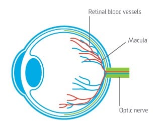
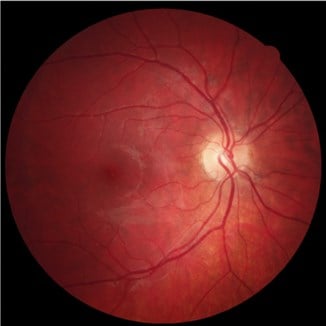
Figure 1: Shows a cross section of the eye and a normal digital retinal image.
|
MACULA |
The part of your retina that is responsible for providing your central sharpest vision. |
|
OPTIC NERVE |
Sends information collected by your retina to your brain where this information is then interpreted. |
|
RETINAL BLOOD VESSELS |
Supply nutrients and oxygen to the cells that make up you retina. |
|
Figure 2: States the function of key structures that form part of your retina. |
|
Case 1 – Good vision doesn’t always mean a healthy retina
Recently, a patient of GMHBA Eye Care had their regular two-yearly eye examination. This patient had excellent vision. At this visit however, a small and very subtle bleed was noticed at the optic nerve of their right eye. This key sign is often an indication of glaucoma. As we were able to compare these new images with previous images, we could confirm that this bleed was a new finding.
This patient was referred to a specialist for glaucoma assessment, and a diagnosis was confirmed. Fortunately this patient is now receiving treatment and further glaucoma progression has been halted and vision loss minimised.
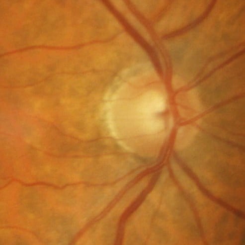
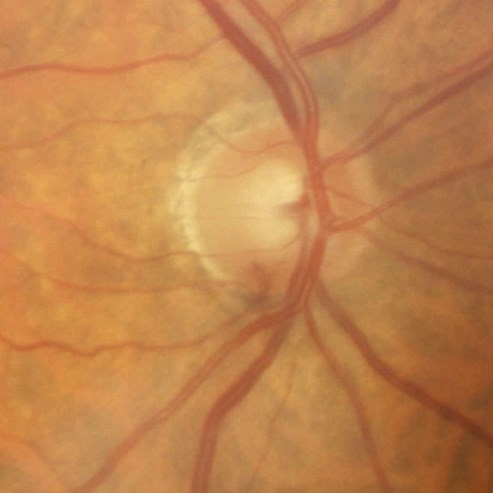
Figure 3: Shows a small bleed at the optic nerve on the image of the right (6 o’clock). This bleed was not noted on an image captured previously (left).
Case 2 – Retina scans are important for anyone with diabetes.
Our second case study was a patient with long-standing diabetes who presented to our practice for the first time due to a sudden blurring of their vision. They had not undergone an eye examination for over 8 years, which is well over the recommended annual review for all people with diabetes. A standard eye health exam of the retina revealed scattered bleeds and patches of fluid leaking from his retinal blood vessels. This is known as diabetic retinopathy.
It wasn’t until digital retinal images were taken the true severity and magnitude of this condition was fully appreciated. See figure 4 below.
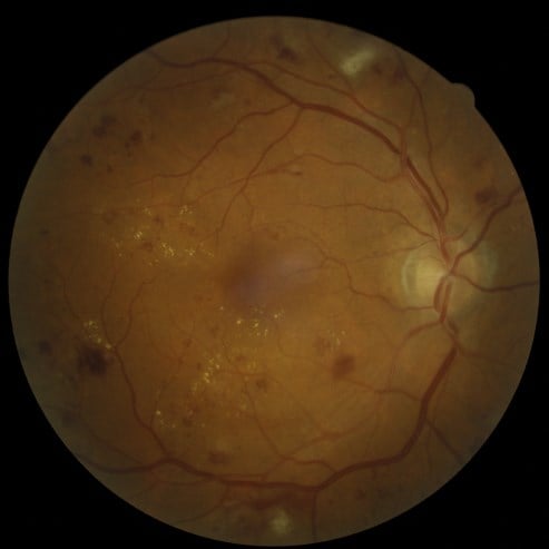
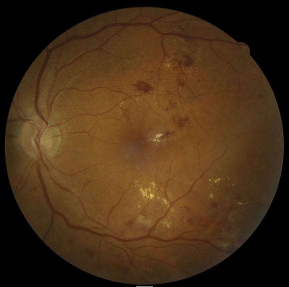
Figure 4: Shows the impact uncontrolled diabetes can have on the retina. These photos show scattered bleeding and fluid (yellow spots) leaking from the blood vessels on both right and left retinas.
Due to the severity of this presentation, this patient was urgently referred to a local ophthalmologist for further assessment and treatment. Their GP was also informed and more aggressive treatment methods were put in place to better control their blood sugar levels.
Case 3 – Retina scans are important for people suffering from high blood pressure.
This case is another example of a patient presenting with a sudden loss of vision. This time, the problem was not diabetes but rather very high blood pressure. As a consequence of this, they had experienced a very large bleed across the lower half of their retina, known as a branch retinal vein occlusion.
Unfortunately for the patient, this bleed had also spread out and completely covered their macula resulting in severe vision loss. This patient was immediately referred to a specialist for treatment at which time their high blood pressure was also addressed.

Figure 5: Shows extensive bleeding across the lower half the retina. Known as a branch retinal vein occlusion.
Case 4 – Regular scans can help you monitor your long term health
The final case I would like to share with you also highlights the importance of regular digital retinal imaging. When this patient attended our practice for the first time an unusually large area of darkened retina was observed and subsequently photographed. As a precaution, this patient was referred to a specialist to rule out any form of malignancy or tumour. Thankfully, this was not the case, but due to having these photos available we are now able to closely monitor this section of retina for any signs of change.

Figure 6: Shows a very large area of darkened retina that requires frequent monitoring.
In summary, digital retinal photography is an extremely effective way to monitor eye health. If you are interested in having your retinas photographed please contact your nearest GMHBA Eye Care practice. Remember you can always ask for a copy to be emailed to you. They make great computer desktop backgrounds!
